
EXAMS · SÃO PAULO
Scrotal ultrasound (Doppler) and individual structures: cost, procedure and preparation
Ultrasound to investigate testicular discomfort, swelling or a suspected varicocele is a quick test that shows local blood flow in real time.

Who performs your exam
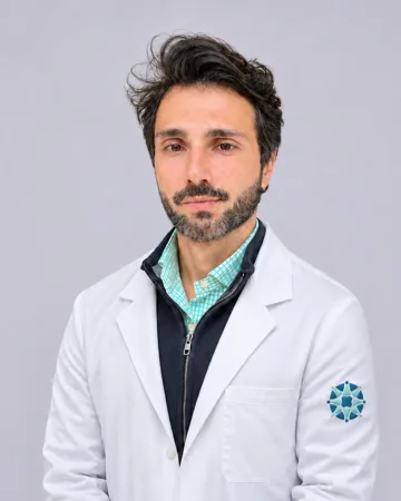
CRM-SP 151361 · RQE 104983
Understand the exam
Scrotal Doppler ultrasound assesses the tissue structure of the testicles and epididymides, including the size, shape and vascular pattern of these structures. In acute scrotal pain, high-frequency color Doppler ultrasound distinguishes acute inflammatory conditions from testicular ischemia or torsion.
The clinic lists the test as Color Doppler ultrasound of an individual organ or structure (scrotum, tendon, muscle). At the clinic, it is performed by Dr. Pedro Moraes.
How it is done
The test uses a high-frequency linear transducer to show detailed testicular anatomy and nearby structures, while Doppler assesses blood flow. When checking for a varicocele, the doctor may ask you to strain your abdomen (Valsalva maneuver) or stand up to see whether the veins dilate.
According to the clinic's records, the test is expected to take 10 minutes and has no preparation listed. The team answers any questions when you book.
When it is recommended
- Detecting and grading varicocele, including during male fertility assessment
- Pain and swelling or enlargement of the scrotum
- Suspected inflammation or infection of the epididymis and testicle (epididymo-orchitis)
- Evaluating epididymal cysts, fluid buildup (hydrocele) or scrotal hernia
- Follow-up after surgery for varicocele or undescended testicles, or assessment of treatment success
Does the exam hurt?
This is a simple, noninvasive test with no exposure to ionizing radiation. If the area is inflamed, it may be tender to the touch.
Cost and health plans
| Exam | Self-pay | Duration | Health plans |
|---|---|---|---|
| Color Doppler of an individual organ or structure (scrotum, tendon, muscle) | Starting at R$ 249.00 (≈ US$ 44, estimated) | 10 minutes | Abas, Bradesco, Mediservice, Omint |
The team confirms the requested option, coverage, and conditions before booking.
Book an examFrequently asked questions
Why does the listed Doppler ultrasound name mention the scrotum, tendon and muscle?
The system follows a procedure schedule that groups Doppler ultrasound of superficial structures under Color Doppler ultrasound of an individual organ or structure (scrotum, tendon, muscle). The doctor examines the exact structure specified on your order.
Do I need to fast or remove hair before scrotal Doppler ultrasound?
No preparation is listed in the clinic's records. Simply maintain your usual hygiene; the team provides instructions when booking if needed.
How long does the exam take?
According to the clinic's records, the expected duration is 10 minutes.
How does Doppler ultrasound detect a varicocele?
A varicocele is a dilation of veins caused by faulty valves in the veins of the pampiniform plexus. Color Doppler ultrasound confirms abnormal venous reflux during the Valsalva maneuver.
Is scrotal Doppler ultrasound covered by my health plan?
Contact the reception team with your doctor's order ready; they will confirm coverage under your health plan.
How long does it take to get the results?
The listing does not specify a report turnaround time. On the day of the test, the team tells you when and how you will receive the result.
Conditions investigated
Where we see patients
References
- Kumar K, Kumari M, Kumar V, et al. Evaluation of Scrotal Pathologies by Ultrasound and Color Doppler. Cureus. 2023.
- Expert Panel on Urological Imaging, Wang CL, Aryal B, et al. ACR Appropriateness Criteria Acute Onset of Scrotal Pain-Without Trauma, Without Antecedent Mass. J Am Coll Radiol. 2019.
- Tyloch JF, Wieczorek AP. Standards for scrotal ultrasonography. J Ultrason. 2016.
- López Juan JA, Palma Maldonado FJ, Cabrera Gómez J, et al. Use of clinical ultrasound in primary care: Scrotal pathology. Semergen. 2024.
- Ebubedike UR, Enukegwu SU, Nwofor AM, et al. Ultrasound evaluation of testicular vein diameter in suspected cases of varicocele: Comparison of measurements in supine and upright positions. Niger J Clin Pract. 2020.
- Freeman S, Bertolotto M, Richenberg J, et al. Ultrasound evaluation of varicoceles: guidelines and recommendations of the European Society of Urogenital Radiology Scrotal and Penile Imaging Working Group (ESUR-SPIWG) for detection, classification, and grading. Eur Radiol. 2020.
- Sosnowska-Sienkiewicz P, Januszkiewicz-Lewandowska D, Mańkowski P. Testicular and scrotal abnormalities in pediatric and adult patients. Pol Przegl Chir. 2023.
- Cao B, Zhang C, Xiao F, et al. Acute epididymo-orchitis complicated by outcomes of either testicular necrosis or complete recovery: Two case reports. Medicine (Baltimore). 2025.
- Spaziani M, Lecis C, Tarantino C, et al. The role of scrotal ultrasonography from infancy to puberty. Andrology. 2021.
- de Grae MNM, Al-Khattab M, Alkadhimi A, et al. A fifteen-year retrospective analysis of varicocele embolization: evaluating success, recurrence rates and embolic agents. CVIR Endovasc. 2025.
- Küçükay F, Tuna HS, et al. Endovascular Embolization of Left-Sided Varicocele: Outcomes by Venographic Anatomy and Coil-Adjunct Strategy. J Vasc Interv Radiol. 2026.
This content is informational and does not replace a consultation.
Updated on